FLT3 single chain antibody
| 【No.】IAB011A | 【Antibody name】 FLT3 single chain antibody |
| 【Antibody subtype】human IgG1 | 【Use】FACS |
| 【Packing specification】100μg/tube | 【Mode of transport】-20°C transportation |
FLT3 belonging Ⅲ receptor tyrosine
kinase family (receptor tyrosine kinase Ⅲ Family, the RTK Ⅲ) . The
members of this family also include c-KIT receptor, platelet-derived
growth factor receptor and c-FMS receptor, etc. The genes of each member
of this family have high homology. FLTS contains 5 extracellular ligand
binding domains composed of immunoglobulin poplar
structure, 1 transmembrane domain, 1 intracellular proximal membrane
domain, 2 kinase domains separated by insertion domains, and
a C- terminal- Structure domain [1] . FLT3 is expressed in
liver, spleen, lymph, brain, placenta and other tissues , and plays an
important role in the proliferation, differentiation and apoptosis
of hematopoietic stem cells, pre- B cells, and dendritic cells. [2] In addition, studies have found that the onset of acute myeloid leukemia is often accompanied by abnormal activation of FLT3 [3] ,
so FLT3 may be used as a potential therapeutic target for acute myeloid
leukemia . This product is an antibody targeting FLT3 , which can be
used for overexpressionFlow cytometric detection of FLT3 recombinant
cell line and detection of recombinant protein activity.
Product name: FLT3 single chain antibody
Reactivity: human
Tags: Fc tag
Subclass: human IgG1
Purification method: affinity chromatography
Storage system: PBS, pH 7.4, 20% glycerol
Storage conditions: -20°C refrigerator, avoid repeated freezing and thawing

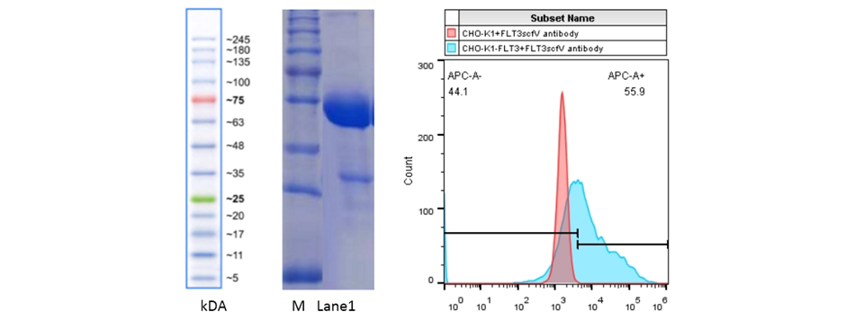
Illustration: Lane M on the left is: protein MW marker, Lane 1 is: 2ug FLT3 single-chain antibody;
The right picture shows the FACS detection of FLT3 antibody binding to FLT3 overexpressing recombinant cell line.
Reference materials:
1. Genomic structure of human Flt3: implications for mutational analysis
2. Flt3-dependent transformation by inactivating c-Cbl mutations in AML
3.Flt3-ITD and tyrosine kinase domain mutants induce-distinct phenotypes in a murine bone marrow transplantation model

